The Iowa Brain Lesion and Neuromodulation Laboratory (BLNL) uses brain imaging and electrical stimulation technology to gain insights into brain-behavior relationships and predict outcomes for patients with a variety of neurological conditions.
Lesion-Behavior Mapping
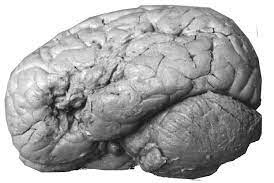
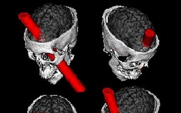
The use of focal brain lesions to infer brain-behavior relationships has been central to cognitive neuroscience since at least the mid-19th century when Paul Broca described the anatomical correlates of expressive aphasia in his famous case study (Figure 1). Around the same time, George Harlow documented the childlike behavior of a formerly mature adult man, Phineas Gage (Figure 2), implicating the prefrontal lobe in personality. These case studies and others like them created foundational knowledge in the field of neuropsychology. Despite advances in functional neuroimaging, the lesion method (or "lesion-deficit mapping") remains critical to cognitive neuroscience and neuropsychology because it allows researchers and clinicians to infer the necessity of different brain structures for human cognition and behavior. In other words, the lesion method allows for "causal" inferences about brain-behavior relationships. This knowledge is essential to the clinical practice of neuropsychology (e.g., in helping to assess the brain regions generating seizure activity in patients with epilepsy). Today, lesion-deficit mapping can be performed using data from hundreds to thousands of patients thanks to advances in neuroimaging registration techniques, the formation of large-scale patient registries, and the creation of statistical pipelines specifically designed for the analysis of lesion data. Our lab uses lesion-behavior mapping to (1) test theories about the structure of cognitive abilities and behavior, and (2) develop methods for predicting cognitive deficits among patients with focal brain lesions (e.g., patients with stroke).

Lesion Network Mapping
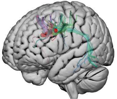
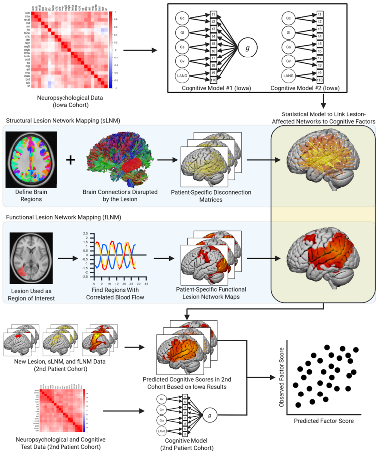
In the mid-20th century, neurologist Norman Geschwind authored a landmark treatise concerning the localization of neurological syndromes to disconnections of long range white matter projections - the highways that allow neurons to communicate with one another. It is now widely recognized that complex thought and behavior likely arise from complex circuitry across distributed anatomical regions of the brain. Lesion network mapping is a statistical approach that integrates the lesion method with techniques from network neuroscience. Lesion-associated networks can be defined both by matter tracts (i.e., bundles of neuronal axons) that overlap with a lesion, and by the brain regions that show correlated blood flow to the affected region (Figure 4). Our lab uses lesion network mapping to (1) infer the shared and unique brain networks that are critical for different aspects of complex cognition and behavior, (2) generate new hypotheses about the structure of the mind, and (3) predict post-lesion deficits.
Intracranial Electrical Stimulation
Direct electrical stimulation of the brain has been a pillar of neurosurgery, and has been a powerful tool for cognitive neuroscience. Patients with drug-resistant epilepsy who are candidates for neurosurgery will often have electrodes placed into the brain to record brain activity related to seizures and identify brain regions necessary for important brain functions like language (which surgeons do their best to avoid to minimize deficits after surgery). However, most electrical stimulation mapping protocols do not involve identifying areas for important cognitive abilities.
Moreover, electrical stimulation mapping of brain functions is typically limited to the brain's cortical surface despite recent lesion studies suggesting that the connections between cortical regions in the brain's "white matter" are more important for explaining cognitive deficits than the brain's cortical surface. Our lab seeks to understand the cognitive and neural mechanisms of important cognitive functions using direct electrical brain stimulation of white matter during specially designed cognitive tests. We also use diffusion weighted imaging-based tractography to visualize the connections affected by the stimulation. Altogether, our work in this area can allow us to learn more about the neuroscience of cognition, and could open the door to clinically useful techniques for mapping cognition to the brain for clinical outcome prediction.